goat igg Search Results


86
Sangon Biotech anti nlrp3 rabbit polyclonal antibody
A qPCR showed increased TLR4 expression in the 216911KO cell line. ** p < 0.01 by student T tests. B The diagram presented two pir-hsa-216911 binding sites in 3ʹUTR of TLR4. C The dual luciferase assay revealed that pir-has-216911 could bind both TLR4 binding sites TLR4S1 and TLR4S2. Plasmids carrying TLR4S1 and TLR4S2 generate a higher Firefly/Renilla Luciferase ratio in the 216911KO cell line, which pir-hsa-216911 was absent. **p < 0.01; ***p < 0.001 by student T tests. D TCGA-LIHC survival analysis found low expression of TLR4 to be unfavorable to the survival of HCC patients. E Western blot analysis revealed pir-hsa-216911 modulating the <t>TLR4/NFκB/NLRP3</t> signaling pathway. TLR4, p-p65, and NLRP3 levels were elevated in the 216911KO cell line, which pir-hsa-216911 was absent. F Western blot analysis revealed pir-hsa-216911 modulating the TLR4/NFκB/NLRP3 signaling pathway through silencing TLR4. After silencing TLR4 with siRNA, pir-hsa-216911 absent could not elevate p-p65, and NLRP3 levels were elevated in the 216911KO cell line.
Anti Nlrp3 Rabbit Polyclonal Antibody, supplied by Sangon Biotech, used in various techniques. Bioz Stars score: 86/100, based on 1 PubMed citations. ZERO BIAS - scores, article reviews, protocol conditions and more
https://www.bioz.com/product/goat+igg/pmc11742400-321-49-56?v=Sangon+Biotech
Average 86 stars, based on 1 article reviews
anti nlrp3 rabbit polyclonal antibody - by Bioz Stars, 2026-08
86/100 stars
  Buy from Supplier

86
Abbkine Inc dylight 549
A qPCR showed increased TLR4 expression in the 216911KO cell line. ** p < 0.01 by student T tests. B The diagram presented two pir-hsa-216911 binding sites in 3ʹUTR of TLR4. C The dual luciferase assay revealed that pir-has-216911 could bind both TLR4 binding sites TLR4S1 and TLR4S2. Plasmids carrying TLR4S1 and TLR4S2 generate a higher Firefly/Renilla Luciferase ratio in the 216911KO cell line, which pir-hsa-216911 was absent. **p < 0.01; ***p < 0.001 by student T tests. D TCGA-LIHC survival analysis found low expression of TLR4 to be unfavorable to the survival of HCC patients. E Western blot analysis revealed pir-hsa-216911 modulating the <t>TLR4/NFκB/NLRP3</t> signaling pathway. TLR4, p-p65, and NLRP3 levels were elevated in the 216911KO cell line, which pir-hsa-216911 was absent. F Western blot analysis revealed pir-hsa-216911 modulating the TLR4/NFκB/NLRP3 signaling pathway through silencing TLR4. After silencing TLR4 with siRNA, pir-hsa-216911 absent could not elevate p-p65, and NLRP3 levels were elevated in the 216911KO cell line.
Dylight 549, supplied by Abbkine Inc, used in various techniques. Bioz Stars score: 86/100, based on 1 PubMed citations. ZERO BIAS - scores, article reviews, protocol conditions and more
https://www.bioz.com/product/goat+igg/pm41814323-65-12-18?v=Abbkine+Inc
Average 86 stars, based on 1 article reviews
dylight 549 - by Bioz Stars, 2026-08
86/100 stars
  Buy from Supplier

86
Servicebio Inc alexa fluor
A qPCR showed increased TLR4 expression in the 216911KO cell line. ** p < 0.01 by student T tests. B The diagram presented two pir-hsa-216911 binding sites in 3ʹUTR of TLR4. C The dual luciferase assay revealed that pir-has-216911 could bind both TLR4 binding sites TLR4S1 and TLR4S2. Plasmids carrying TLR4S1 and TLR4S2 generate a higher Firefly/Renilla Luciferase ratio in the 216911KO cell line, which pir-hsa-216911 was absent. **p < 0.01; ***p < 0.001 by student T tests. D TCGA-LIHC survival analysis found low expression of TLR4 to be unfavorable to the survival of HCC patients. E Western blot analysis revealed pir-hsa-216911 modulating the <t>TLR4/NFκB/NLRP3</t> signaling pathway. TLR4, p-p65, and NLRP3 levels were elevated in the 216911KO cell line, which pir-hsa-216911 was absent. F Western blot analysis revealed pir-hsa-216911 modulating the TLR4/NFκB/NLRP3 signaling pathway through silencing TLR4. After silencing TLR4 with siRNA, pir-hsa-216911 absent could not elevate p-p65, and NLRP3 levels were elevated in the 216911KO cell line.
Alexa Fluor, supplied by Servicebio Inc, used in various techniques. Bioz Stars score: 86/100, based on 1 PubMed citations. ZERO BIAS - scores, article reviews, protocol conditions and more
https://www.bioz.com/product/goat+igg/pmc12874108-280-108-115?v=Servicebio+Inc
Average 86 stars, based on 1 article reviews
alexa fluor - by Bioz Stars, 2026-08
86/100 stars
  Buy from Supplier

86
Abbkine Inc goat anti rabbit igg
A qPCR showed increased TLR4 expression in the 216911KO cell line. ** p < 0.01 by student T tests. B The diagram presented two pir-hsa-216911 binding sites in 3ʹUTR of TLR4. C The dual luciferase assay revealed that pir-has-216911 could bind both TLR4 binding sites TLR4S1 and TLR4S2. Plasmids carrying TLR4S1 and TLR4S2 generate a higher Firefly/Renilla Luciferase ratio in the 216911KO cell line, which pir-hsa-216911 was absent. **p < 0.01; ***p < 0.001 by student T tests. D TCGA-LIHC survival analysis found low expression of TLR4 to be unfavorable to the survival of HCC patients. E Western blot analysis revealed pir-hsa-216911 modulating the <t>TLR4/NFκB/NLRP3</t> signaling pathway. TLR4, p-p65, and NLRP3 levels were elevated in the 216911KO cell line, which pir-hsa-216911 was absent. F Western blot analysis revealed pir-hsa-216911 modulating the TLR4/NFκB/NLRP3 signaling pathway through silencing TLR4. After silencing TLR4 with siRNA, pir-hsa-216911 absent could not elevate p-p65, and NLRP3 levels were elevated in the 216911KO cell line.
Goat Anti Rabbit Igg, supplied by Abbkine Inc, used in various techniques. Bioz Stars score: 86/100, based on 1 PubMed citations. ZERO BIAS - scores, article reviews, protocol conditions and more
https://www.bioz.com/product/goat+igg/pmc12392501-106-15-22?v=Abbkine+Inc
Average 86 stars, based on 1 article reviews
goat anti rabbit igg - by Bioz Stars, 2026-08
86/100 stars
  Buy from Supplier

86
Abbkine Inc dylight 800
A qPCR showed increased TLR4 expression in the 216911KO cell line. ** p < 0.01 by student T tests. B The diagram presented two pir-hsa-216911 binding sites in 3ʹUTR of TLR4. C The dual luciferase assay revealed that pir-has-216911 could bind both TLR4 binding sites TLR4S1 and TLR4S2. Plasmids carrying TLR4S1 and TLR4S2 generate a higher Firefly/Renilla Luciferase ratio in the 216911KO cell line, which pir-hsa-216911 was absent. **p < 0.01; ***p < 0.001 by student T tests. D TCGA-LIHC survival analysis found low expression of TLR4 to be unfavorable to the survival of HCC patients. E Western blot analysis revealed pir-hsa-216911 modulating the <t>TLR4/NFκB/NLRP3</t> signaling pathway. TLR4, p-p65, and NLRP3 levels were elevated in the 216911KO cell line, which pir-hsa-216911 was absent. F Western blot analysis revealed pir-hsa-216911 modulating the TLR4/NFκB/NLRP3 signaling pathway through silencing TLR4. After silencing TLR4 with siRNA, pir-hsa-216911 absent could not elevate p-p65, and NLRP3 levels were elevated in the 216911KO cell line.
Dylight 800, supplied by Abbkine Inc, used in various techniques. Bioz Stars score: 86/100, based on 1 PubMed citations. ZERO BIAS - scores, article reviews, protocol conditions and more
https://www.bioz.com/product/goat+igg/pm41139043-151-16-24?v=Abbkine+Inc
Average 86 stars, based on 1 article reviews
dylight 800 - by Bioz Stars, 2026-08
86/100 stars
  Buy from Supplier

86
Wuhan Sanying Biotechnology hrp conjugated goat
A qPCR showed increased TLR4 expression in the 216911KO cell line. ** p < 0.01 by student T tests. B The diagram presented two pir-hsa-216911 binding sites in 3ʹUTR of TLR4. C The dual luciferase assay revealed that pir-has-216911 could bind both TLR4 binding sites TLR4S1 and TLR4S2. Plasmids carrying TLR4S1 and TLR4S2 generate a higher Firefly/Renilla Luciferase ratio in the 216911KO cell line, which pir-hsa-216911 was absent. **p < 0.01; ***p < 0.001 by student T tests. D TCGA-LIHC survival analysis found low expression of TLR4 to be unfavorable to the survival of HCC patients. E Western blot analysis revealed pir-hsa-216911 modulating the <t>TLR4/NFκB/NLRP3</t> signaling pathway. TLR4, p-p65, and NLRP3 levels were elevated in the 216911KO cell line, which pir-hsa-216911 was absent. F Western blot analysis revealed pir-hsa-216911 modulating the TLR4/NFκB/NLRP3 signaling pathway through silencing TLR4. After silencing TLR4 with siRNA, pir-hsa-216911 absent could not elevate p-p65, and NLRP3 levels were elevated in the 216911KO cell line.
Hrp Conjugated Goat, supplied by Wuhan Sanying Biotechnology, used in various techniques. Bioz Stars score: 86/100, based on 1 PubMed citations. ZERO BIAS - scores, article reviews, protocol conditions and more
https://www.bioz.com/product/goat+igg/pm41456437-118-6-15?v=Wuhan+Sanying+Biotechnology
Average 86 stars, based on 1 article reviews
hrp conjugated goat - by Bioz Stars, 2026-08
86/100 stars
  Buy from Supplier

94
R&D Systems goat anti rat il 6mab
A qPCR showed increased TLR4 expression in the 216911KO cell line. ** p < 0.01 by student T tests. B The diagram presented two pir-hsa-216911 binding sites in 3ʹUTR of TLR4. C The dual luciferase assay revealed that pir-has-216911 could bind both TLR4 binding sites TLR4S1 and TLR4S2. Plasmids carrying TLR4S1 and TLR4S2 generate a higher Firefly/Renilla Luciferase ratio in the 216911KO cell line, which pir-hsa-216911 was absent. **p < 0.01; ***p < 0.001 by student T tests. D TCGA-LIHC survival analysis found low expression of TLR4 to be unfavorable to the survival of HCC patients. E Western blot analysis revealed pir-hsa-216911 modulating the <t>TLR4/NFκB/NLRP3</t> signaling pathway. TLR4, p-p65, and NLRP3 levels were elevated in the 216911KO cell line, which pir-hsa-216911 was absent. F Western blot analysis revealed pir-hsa-216911 modulating the TLR4/NFκB/NLRP3 signaling pathway through silencing TLR4. After silencing TLR4 with siRNA, pir-hsa-216911 absent could not elevate p-p65, and NLRP3 levels were elevated in the 216911KO cell line.
Goat Anti Rat Il 6mab, supplied by R&D Systems, used in various techniques. Bioz Stars score: 94/100, based on 1 PubMed citations. ZERO BIAS - scores, article reviews, protocol conditions and more
https://www.bioz.com/product/goat+igg/pmc08716408-34-6-8?v=R%26D+Systems
Average 94 stars, based on 1 article reviews
goat anti rat il 6mab - by Bioz Stars, 2026-08
94/100 stars
  Buy from Supplier

95
Boster Bio anti mouse igg
A qPCR showed increased TLR4 expression in the 216911KO cell line. ** p < 0.01 by student T tests. B The diagram presented two pir-hsa-216911 binding sites in 3ʹUTR of TLR4. C The dual luciferase assay revealed that pir-has-216911 could bind both TLR4 binding sites TLR4S1 and TLR4S2. Plasmids carrying TLR4S1 and TLR4S2 generate a higher Firefly/Renilla Luciferase ratio in the 216911KO cell line, which pir-hsa-216911 was absent. **p < 0.01; ***p < 0.001 by student T tests. D TCGA-LIHC survival analysis found low expression of TLR4 to be unfavorable to the survival of HCC patients. E Western blot analysis revealed pir-hsa-216911 modulating the <t>TLR4/NFκB/NLRP3</t> signaling pathway. TLR4, p-p65, and NLRP3 levels were elevated in the 216911KO cell line, which pir-hsa-216911 was absent. F Western blot analysis revealed pir-hsa-216911 modulating the TLR4/NFκB/NLRP3 signaling pathway through silencing TLR4. After silencing TLR4 with siRNA, pir-hsa-216911 absent could not elevate p-p65, and NLRP3 levels were elevated in the 216911KO cell line.
Anti Mouse Igg, supplied by Boster Bio, used in various techniques. Bioz Stars score: 95/100, based on 1 PubMed citations. ZERO BIAS - scores, article reviews, protocol conditions and more
https://www.bioz.com/product/goat+igg/pmc12857345-16-16-22?v=Boster+Bio
Average 95 stars, based on 1 article reviews
anti mouse igg - by Bioz Stars, 2026-08
95/100 stars
  Buy from Supplier

96
Boster Bio goat anti rabbit igg
A qPCR showed increased TLR4 expression in the 216911KO cell line. ** p < 0.01 by student T tests. B The diagram presented two pir-hsa-216911 binding sites in 3ʹUTR of TLR4. C The dual luciferase assay revealed that pir-has-216911 could bind both TLR4 binding sites TLR4S1 and TLR4S2. Plasmids carrying TLR4S1 and TLR4S2 generate a higher Firefly/Renilla Luciferase ratio in the 216911KO cell line, which pir-hsa-216911 was absent. **p < 0.01; ***p < 0.001 by student T tests. D TCGA-LIHC survival analysis found low expression of TLR4 to be unfavorable to the survival of HCC patients. E Western blot analysis revealed pir-hsa-216911 modulating the <t>TLR4/NFκB/NLRP3</t> signaling pathway. TLR4, p-p65, and NLRP3 levels were elevated in the 216911KO cell line, which pir-hsa-216911 was absent. F Western blot analysis revealed pir-hsa-216911 modulating the TLR4/NFκB/NLRP3 signaling pathway through silencing TLR4. After silencing TLR4 with siRNA, pir-hsa-216911 absent could not elevate p-p65, and NLRP3 levels were elevated in the 216911KO cell line.
Goat Anti Rabbit Igg, supplied by Boster Bio, used in various techniques. Bioz Stars score: 96/100, based on 1 PubMed citations. ZERO BIAS - scores, article reviews, protocol conditions and more
https://www.bioz.com/product/goat+igg/pmc12967463-7-0-11?v=Boster+Bio
Average 96 stars, based on 1 article reviews
goat anti rabbit igg - by Bioz Stars, 2026-08
96/100 stars
  Buy from Supplier

99
R&D Systems anti goat igg hrp
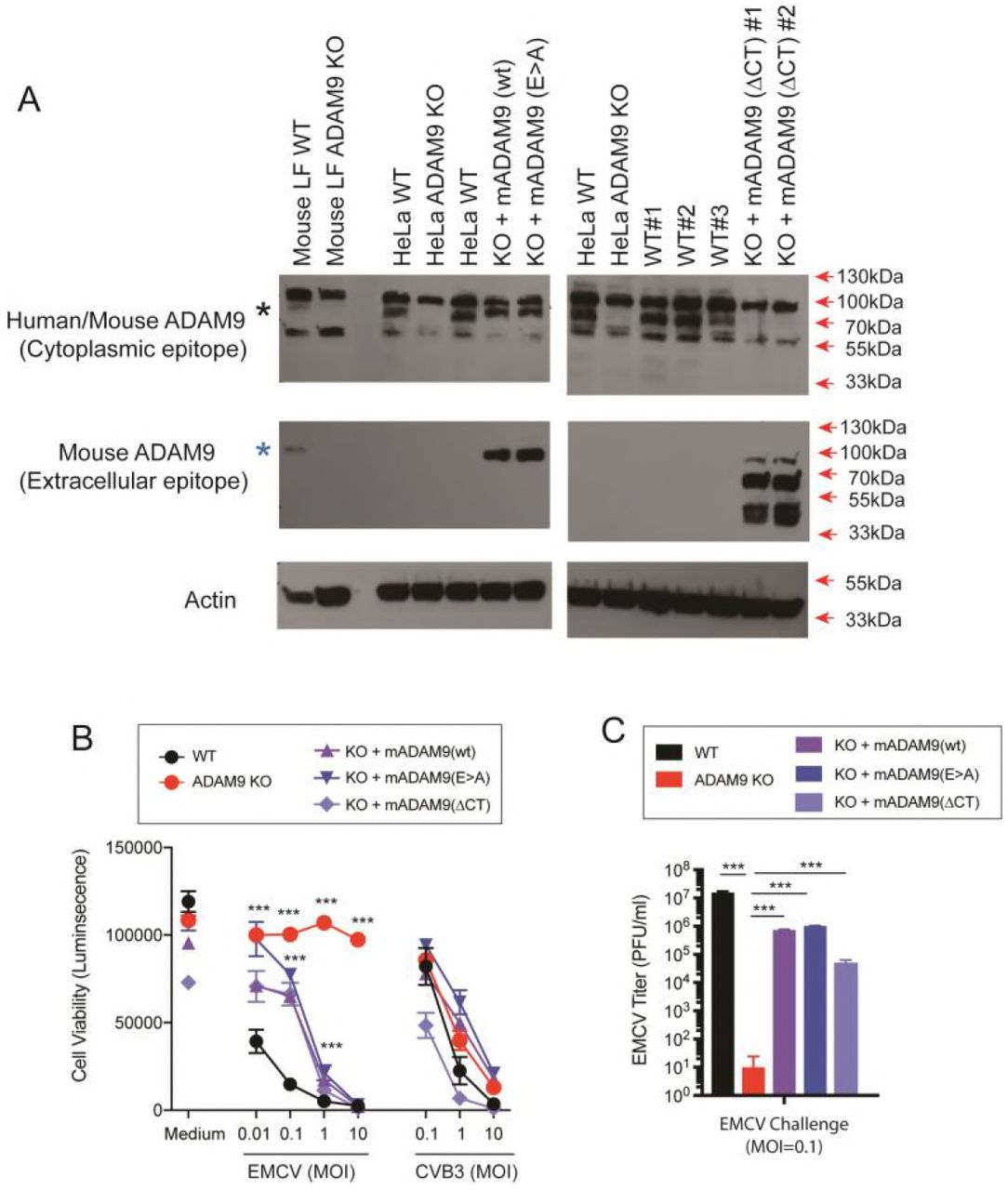
WT and ADAM9 KO HeLa cells were transduced with retroviral vectors with wild-type (WT) murine ADAM9 (mADAM9), catalytically inactive mutant ADAM9 (E>A), cytoplasmic tail deleted (ΔCT) ADAM9 constructs, or GFP control vectors. Transduced cells were selected and cloned by limiting dilution, and expression of mADAM9 was confirmed by western blot analysis. (A) WT, KO and rescue cell lysates were prepared in RIPA buffer and run on 12% SDS-PAGE gels and transferred to PVDF membranes. Membranes were blocked with <t>3%</t> <t>BSA</t> and incubated with ADAM9 antibodies. Upper panel, rabbit anti-human ADAM9 (Cell signaling #2099) that detects an epitope in the intracellular domain of human ADAM9 and cross-reacts with mouse ADAM9. Middle panel, goat anti-mouse ADAM9 (R&D systems AF949) that detects the extracellular domain of murine ADAM9, but not human ADAM9. Lower panel, anti-Actin (Santa Cruz sc-1616) which detects both human and murine ϟ-Actin. Bands were visualized using <t>HRP</t> and ECL reagent. Upper panel: WT but not KO cells expressed human ADAM9. Middle panel: Rescue but not KO cells expressed murine ADAM9. Lower panel: Actin loading control. (B) WT clones, ADAM9 KO clones and rescued ADAM9-expressing clones were infected with EMCV or CVB3 at varying MOI and incubated at 37 °C for 24 h. Viability of EMCV-infected and CVB3-infected clones was measured by CellGlo ATP luminescence. (C) EMCV replication was quantified in infected culture supernatants by plaque assay using BHK-21 cells. Neither the functional sequence of the ADAM9 metalloproteinase domain nor the cytoplasmic tail are required for EMCV infection. ***, P <0.0001, KO vs. WT; KO vs. rescue.
Anti Goat Igg Hrp, supplied by R&D Systems, used in various techniques. Bioz Stars score: 99/100, based on 1 PubMed citations. ZERO BIAS - scores, article reviews, protocol conditions and more
https://www.bioz.com/product/goat+igg/bio_rxiv__491068-209-4-9?v=R%26D+Systems
Average 99 stars, based on 1 article reviews
anti goat igg hrp - by Bioz Stars, 2026-08
99/100 stars
  Buy from Supplier

99
Novus Biologicals antibody conjugated to goat anti mouse hrp
WT and ADAM9 KO HeLa cells were transduced with retroviral vectors with wild-type (WT) murine ADAM9 (mADAM9), catalytically inactive mutant ADAM9 (E>A), cytoplasmic tail deleted (ΔCT) ADAM9 constructs, or GFP control vectors. Transduced cells were selected and cloned by limiting dilution, and expression of mADAM9 was confirmed by western blot analysis. (A) WT, KO and rescue cell lysates were prepared in RIPA buffer and run on 12% SDS-PAGE gels and transferred to PVDF membranes. Membranes were blocked with <t>3%</t> <t>BSA</t> and incubated with ADAM9 antibodies. Upper panel, rabbit anti-human ADAM9 (Cell signaling #2099) that detects an epitope in the intracellular domain of human ADAM9 and cross-reacts with mouse ADAM9. Middle panel, goat anti-mouse ADAM9 (R&D systems AF949) that detects the extracellular domain of murine ADAM9, but not human ADAM9. Lower panel, anti-Actin (Santa Cruz sc-1616) which detects both human and murine ϟ-Actin. Bands were visualized using <t>HRP</t> and ECL reagent. Upper panel: WT but not KO cells expressed human ADAM9. Middle panel: Rescue but not KO cells expressed murine ADAM9. Lower panel: Actin loading control. (B) WT clones, ADAM9 KO clones and rescued ADAM9-expressing clones were infected with EMCV or CVB3 at varying MOI and incubated at 37 °C for 24 h. Viability of EMCV-infected and CVB3-infected clones was measured by CellGlo ATP luminescence. (C) EMCV replication was quantified in infected culture supernatants by plaque assay using BHK-21 cells. Neither the functional sequence of the ADAM9 metalloproteinase domain nor the cytoplasmic tail are required for EMCV infection. ***, P <0.0001, KO vs. WT; KO vs. rescue.
Antibody Conjugated To Goat Anti Mouse Hrp, supplied by Novus Biologicals, used in various techniques. Bioz Stars score: 99/100, based on 1 PubMed citations. ZERO BIAS - scores, article reviews, protocol conditions and more
https://www.bioz.com/product/goat+igg/bio_rxiv__2021__01__06__425530-309-19-26?v=Novus+Biologicals
Average 99 stars, based on 1 article reviews
antibody conjugated to goat anti mouse hrp - by Bioz Stars, 2026-08
99/100 stars
  Buy from Supplier

96
R&D Systems control goat igg antibody
Figure 3. Induction of HO-1 by Hb:Hp via a CD163 receptor IL-10 autocrine mechanism. A, Representative HO-1 and HO-2 immunoblots are shown from in vitro differentiated day 7 macrophages stimulated with Hb, Hp, Hb:Hp, or Hb:Hp plus blocking anti–IL-10, <t>IgG</t> <t>control,</t> or RM3/1 <t>antibody.</t> Induction of the HO-1 isomer was established relative to con- stitutive expression of the HO-2 isomer by probing blots with polyclonal antibody against HO-1, followed by stripping and reprobing with anti–HO-2. B, Lysates in A were subjected to quantification of intracellular HO-1 levels by HO-1–spe- cific ELISA and normalized with respect to total cell protein. Values shown repre- sent meanSEM. Results are represen- tative of 3 similar experiments.
Control Goat Igg Antibody, supplied by R&D Systems, used in various techniques. Bioz Stars score: 96/100, based on 1 PubMed citations. ZERO BIAS - scores, article reviews, protocol conditions and more
https://www.bioz.com/product/goat+igg/10__1161_slash_01__res__0000109414__78907__f9-35-7-14?v=R%26D+Systems
Average 96 stars, based on 1 article reviews
control goat igg antibody - by Bioz Stars, 2026-08
96/100 stars
  Buy from Supplier

Image Search Results


A qPCR showed increased TLR4 expression in the 216911KO cell line. ** p < 0.01 by student T tests. B The diagram presented two pir-hsa-216911 binding sites in 3ʹUTR of TLR4. C The dual luciferase assay revealed that pir-has-216911 could bind both TLR4 binding sites TLR4S1 and TLR4S2. Plasmids carrying TLR4S1 and TLR4S2 generate a higher Firefly/Renilla Luciferase ratio in the 216911KO cell line, which pir-hsa-216911 was absent. **p < 0.01; ***p < 0.001 by student T tests. D TCGA-LIHC survival analysis found low expression of TLR4 to be unfavorable to the survival of HCC patients. E Western blot analysis revealed pir-hsa-216911 modulating the TLR4/NFκB/NLRP3 signaling pathway. TLR4, p-p65, and NLRP3 levels were elevated in the 216911KO cell line, which pir-hsa-216911 was absent. F Western blot analysis revealed pir-hsa-216911 modulating the TLR4/NFκB/NLRP3 signaling pathway through silencing TLR4. After silencing TLR4 with siRNA, pir-hsa-216911 absent could not elevate p-p65, and NLRP3 levels were elevated in the 216911KO cell line.

Journal: Cell Death Discovery

Article Title: pir-hsa-216911 inhibit pyroptosis in hepatocellular carcinoma by suppressing TLR4 initiated GSDMD activation

doi: 10.1038/s41420-024-02285-9

Figure Lengend Snippet: A qPCR showed increased TLR4 expression in the 216911KO cell line. ** p < 0.01 by student T tests. B The diagram presented two pir-hsa-216911 binding sites in 3ʹUTR of TLR4. C The dual luciferase assay revealed that pir-has-216911 could bind both TLR4 binding sites TLR4S1 and TLR4S2. Plasmids carrying TLR4S1 and TLR4S2 generate a higher Firefly/Renilla Luciferase ratio in the 216911KO cell line, which pir-hsa-216911 was absent. **p < 0.01; ***p < 0.001 by student T tests. D TCGA-LIHC survival analysis found low expression of TLR4 to be unfavorable to the survival of HCC patients. E Western blot analysis revealed pir-hsa-216911 modulating the TLR4/NFκB/NLRP3 signaling pathway. TLR4, p-p65, and NLRP3 levels were elevated in the 216911KO cell line, which pir-hsa-216911 was absent. F Western blot analysis revealed pir-hsa-216911 modulating the TLR4/NFκB/NLRP3 signaling pathway through silencing TLR4. After silencing TLR4 with siRNA, pir-hsa-216911 absent could not elevate p-p65, and NLRP3 levels were elevated in the 216911KO cell line.

Article Snippet: The antibodies used in this study were Cleaved Caspase-1 Rabbit mAb (4199, Cell Signaling Technology, USA), Cleaved Gasdermin D Rabbit mAb (1:2000 dilute; 36425 Cell Signaling Technology, USA), anti-TLR4 rabbit polyclonal antibody (1:500 dilute; D121751, Sangon Biotech, China), anti-RELA (Phospho-Ser536) rabbit polyclonal antibody (1:500 dilute; D155006, Sangon Biotech, China), anti-NLRP3 rabbit polyclonal antibody (1:250 dilute; D120143, Sangon Biotech, China), anti-ACTB rabbit polyclonal antibody (1:6000 dilute; D110001, Sangon Biotech, China), anti-GAPDH rabbit polyclonal antibody (1:4000 dilute; D110016, Sangon Biotech, China), and HRP-conjugated goat anti-rabbit IgG (1:6000 dilute; D110058, Sangon Biotech, China).

Techniques: Expressing, Binding Assay, Luciferase, Western Blot

A The diagram presented the xenograft modeling process. B Euthanized mouse to display the subcutaneous xenografted tumors. C 216911KO group resulted in much smaller tumors in size compared to the control group. D 216911KO group resulted in much smaller tumors in weight compared to the control group. The histogram represents the mean tumor weight of each group. ***p < 0.001 by student T tests. E Western blot analysis revealed pir-hsa-216911 modulating the TLR4/NFκB/NLRP3 signaling pathway in vivo. Both TLR4 and p-p65 levels were elevated in tumors formed in the 216911KO group compared to the control group.

Journal: Cell Death Discovery

Article Title: pir-hsa-216911 inhibit pyroptosis in hepatocellular carcinoma by suppressing TLR4 initiated GSDMD activation

doi: 10.1038/s41420-024-02285-9

Figure Lengend Snippet: A The diagram presented the xenograft modeling process. B Euthanized mouse to display the subcutaneous xenografted tumors. C 216911KO group resulted in much smaller tumors in size compared to the control group. D 216911KO group resulted in much smaller tumors in weight compared to the control group. The histogram represents the mean tumor weight of each group. ***p < 0.001 by student T tests. E Western blot analysis revealed pir-hsa-216911 modulating the TLR4/NFκB/NLRP3 signaling pathway in vivo. Both TLR4 and p-p65 levels were elevated in tumors formed in the 216911KO group compared to the control group.

Article Snippet: The antibodies used in this study were Cleaved Caspase-1 Rabbit mAb (4199, Cell Signaling Technology, USA), Cleaved Gasdermin D Rabbit mAb (1:2000 dilute; 36425 Cell Signaling Technology, USA), anti-TLR4 rabbit polyclonal antibody (1:500 dilute; D121751, Sangon Biotech, China), anti-RELA (Phospho-Ser536) rabbit polyclonal antibody (1:500 dilute; D155006, Sangon Biotech, China), anti-NLRP3 rabbit polyclonal antibody (1:250 dilute; D120143, Sangon Biotech, China), anti-ACTB rabbit polyclonal antibody (1:6000 dilute; D110001, Sangon Biotech, China), anti-GAPDH rabbit polyclonal antibody (1:4000 dilute; D110016, Sangon Biotech, China), and HRP-conjugated goat anti-rabbit IgG (1:6000 dilute; D110058, Sangon Biotech, China).

Techniques: Control, Western Blot, In Vivo

WT and ADAM9 KO HeLa cells were transduced with retroviral vectors with wild-type (WT) murine ADAM9 (mADAM9), catalytically inactive mutant ADAM9 (E>A), cytoplasmic tail deleted (ΔCT) ADAM9 constructs, or GFP control vectors. Transduced cells were selected and cloned by limiting dilution, and expression of mADAM9 was confirmed by western blot analysis. (A) WT, KO and rescue cell lysates were prepared in RIPA buffer and run on 12% SDS-PAGE gels and transferred to PVDF membranes. Membranes were blocked with 3% BSA and incubated with ADAM9 antibodies. Upper panel, rabbit anti-human ADAM9 (Cell signaling #2099) that detects an epitope in the intracellular domain of human ADAM9 and cross-reacts with mouse ADAM9. Middle panel, goat anti-mouse ADAM9 (R&D systems AF949) that detects the extracellular domain of murine ADAM9, but not human ADAM9. Lower panel, anti-Actin (Santa Cruz sc-1616) which detects both human and murine ϟ-Actin. Bands were visualized using HRP and ECL reagent. Upper panel: WT but not KO cells expressed human ADAM9. Middle panel: Rescue but not KO cells expressed murine ADAM9. Lower panel: Actin loading control. (B) WT clones, ADAM9 KO clones and rescued ADAM9-expressing clones were infected with EMCV or CVB3 at varying MOI and incubated at 37 °C for 24 h. Viability of EMCV-infected and CVB3-infected clones was measured by CellGlo ATP luminescence. (C) EMCV replication was quantified in infected culture supernatants by plaque assay using BHK-21 cells. Neither the functional sequence of the ADAM9 metalloproteinase domain nor the cytoplasmic tail are required for EMCV infection. ***, P <0.0001, KO vs. WT; KO vs. rescue.

Journal: bioRxiv

Article Title: Identification of A Disintegrin and Metalloproteinase 9 domain (ADAM9) required in the early stages of encephalomyocarditis virus infection

doi: 10.1101/491068

Figure Lengend Snippet: WT and ADAM9 KO HeLa cells were transduced with retroviral vectors with wild-type (WT) murine ADAM9 (mADAM9), catalytically inactive mutant ADAM9 (E>A), cytoplasmic tail deleted (ΔCT) ADAM9 constructs, or GFP control vectors. Transduced cells were selected and cloned by limiting dilution, and expression of mADAM9 was confirmed by western blot analysis. (A) WT, KO and rescue cell lysates were prepared in RIPA buffer and run on 12% SDS-PAGE gels and transferred to PVDF membranes. Membranes were blocked with 3% BSA and incubated with ADAM9 antibodies. Upper panel, rabbit anti-human ADAM9 (Cell signaling #2099) that detects an epitope in the intracellular domain of human ADAM9 and cross-reacts with mouse ADAM9. Middle panel, goat anti-mouse ADAM9 (R&D systems AF949) that detects the extracellular domain of murine ADAM9, but not human ADAM9. Lower panel, anti-Actin (Santa Cruz sc-1616) which detects both human and murine ϟ-Actin. Bands were visualized using HRP and ECL reagent. Upper panel: WT but not KO cells expressed human ADAM9. Middle panel: Rescue but not KO cells expressed murine ADAM9. Lower panel: Actin loading control. (B) WT clones, ADAM9 KO clones and rescued ADAM9-expressing clones were infected with EMCV or CVB3 at varying MOI and incubated at 37 °C for 24 h. Viability of EMCV-infected and CVB3-infected clones was measured by CellGlo ATP luminescence. (C) EMCV replication was quantified in infected culture supernatants by plaque assay using BHK-21 cells. Neither the functional sequence of the ADAM9 metalloproteinase domain nor the cytoplasmic tail are required for EMCV infection. ***, P <0.0001, KO vs. WT; KO vs. rescue.

Article Snippet: 3% BSA blocking buffer), anti-goat IgG HRP (1:2000 dilution, R&D Systems HAF109), and anti-rabbit IgG HRP (1:5000 dilution Vector Labs PI1000).

Techniques: Transduction, Mutagenesis, Construct, Clone Assay, Expressing, Western Blot, SDS Page, Incubation, Infection, Plaque Assay, Functional Assay, Sequencing

Figure 3. Induction of HO-1 by Hb:Hp via a CD163 receptor IL-10 autocrine mechanism. A, Representative HO-1 and HO-2 immunoblots are shown from in vitro differentiated day 7 macrophages stimulated with Hb, Hp, Hb:Hp, or Hb:Hp plus blocking anti–IL-10, IgG control, or RM3/1 antibody. Induction of the HO-1 isomer was established relative to con- stitutive expression of the HO-2 isomer by probing blots with polyclonal antibody against HO-1, followed by stripping and reprobing with anti–HO-2. B, Lysates in A were subjected to quantification of intracellular HO-1 levels by HO-1–spe- cific ELISA and normalized with respect to total cell protein. Values shown repre- sent meanSEM. Results are represen- tative of 3 similar experiments.

Journal: Circulation Research

Article Title: Hemoglobin Scavenger Receptor CD163 Mediates Interleukin-10 Release and Heme Oxygenase-1 Synthesis

doi: 10.1161/01.res.0000109414.78907.f9

Figure Lengend Snippet: Figure 3. Induction of HO-1 by Hb:Hp via a CD163 receptor IL-10 autocrine mechanism. A, Representative HO-1 and HO-2 immunoblots are shown from in vitro differentiated day 7 macrophages stimulated with Hb, Hp, Hb:Hp, or Hb:Hp plus blocking anti–IL-10, IgG control, or RM3/1 antibody. Induction of the HO-1 isomer was established relative to con- stitutive expression of the HO-2 isomer by probing blots with polyclonal antibody against HO-1, followed by stripping and reprobing with anti–HO-2. B, Lysates in A were subjected to quantification of intracellular HO-1 levels by HO-1–spe- cific ELISA and normalized with respect to total cell protein. Values shown repre- sent meanSEM. Results are represen- tative of 3 similar experiments.

Article Snippet: Neutralizing anti- human IL-10 polyclonal antibody and control goat IgG antibody were purchased from R&D Systems (Abingdon, UK).

Techniques: Western Blot, In Vitro, Blocking Assay, Control, Expressing, Stripping Membranes, Enzyme-linked Immunosorbent Assay